A split-second is all it takes for life to change. A fall, a fire, an auto accident; a trip to the hospital. Traumatic injuries require immediate care to stabilize the patient. After addressing the initial trauma, facial reconstruction surgery can offer a new beginning. We are proud to offer the full spectrum of post-traumatic facial reconstructive services to patients in the Denver area.
Dial 303-839-1616 today to speak with Dr. Robert G. Fante about whether you are a candidate for post-traumatic facial reconstructive!
Patient Testimonial
"I have had the privilege of being a patient Dr Fante's for the past 18 months. He and his staff work tirelessly to provide me with the very best care. The kindness and compassion that I have received sets the bar very high for all my other caregivers. Not only do I get the best medical care but there is such phenomenal emotional support that couples with that care. I consulted with other surgeons and I'm so grateful that I was referred to Fante Eye & Face Centre. I live in Boulder and it is well worth the drive. Oftentimes I can be seen in Boulder or in Broomfield this is done by Dr Fante and staff so that his patients do not have to travel so far. It is difficult in this complex world of insurance and networks of doctors to find such a wonderful place to receive the very best care. My family and I are very grateful. Edna Benson - Boulder Colorado"
What is Complex Post-traumatic Facial Reconstruction?
Reconstruction is a critical aspect of post-trauma care for many children and adults. Complex trauma to the face and head poses extraordinary dilemmas, as numerous structures may have been affected in the initial injury. It is nearly impossible to predict how nerves, muscles, bones, and soft tissue will heal, though this is precisely what experienced oculofacial plastic surgeons try to do.
Observing the face after initial healing has occurred, a surgeon anticipates future healing and the potential for deformity, asymmetry, atrophy, and scarring. Post-traumatic facial reconstruction is performed to mitigate these consequences while preserving nerve and sensory function in bones, muscles, blood vessels, and skin. As much as possible, surgery aims to salvage fundamental structure.
Facial reconstruction is sometimes accomplished in the first weeks after trauma or destruction from cancer. Complex initial repair of bone fractures and soft tissue injuries is usually performed as soon as the person is stable enough to undergo surgery. Repair of broken facial bones may require titanium plates and screws, or implants such as porous polyethylene. Lacerated tear ducts may require microscopic re-approximation and temporary silicone stenting. Tendons, ligaments, muscles and skin each may require meticulous repair.
But sometimes additional surgery may be required months or years later as tissue healing and the ongoing evolution of structural relationships between facial components develops. For example, after trauma, eye sockets sometimes lose fat volume or develop structural weakness that causes the eyeball to sink down and lose mobility, causing deformity and double vision.
How is Facial Reconstruction Performed?
Facial reconstruction is a delicate process that may involve more than one surgery. The degree of remodeling depends on the severity of the injury and the body’s natural healing response. Dr. Fante uses innovative methods of bone and tissue repair to recreate facial features to look as natural as possible. Shape and symmetry are restored using the patient’s own tissues if this can be achieved. Otherwise, base structure may be reinstated utilizing appropriate facial implants, grafts, titanium, and other materials.
Surgical techniques may include endoscopic nasal surgery and tissue grafting as necessary (bone, skin, and cartilage). Tissue may be rearranged using flap reconstruction, and scars are corrected as much as possible to restore normal appearance.
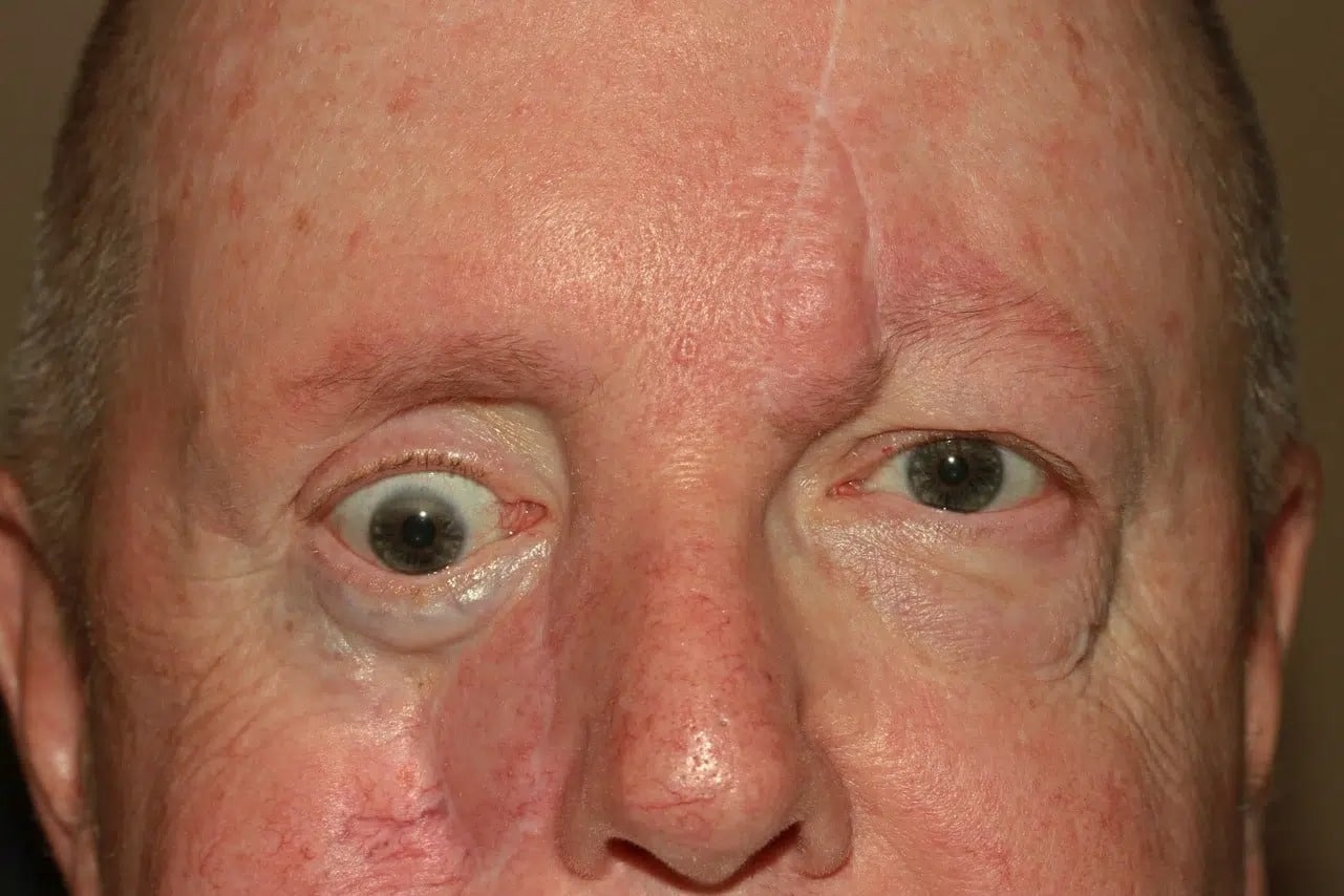
This man had been in a motor vehicle accident 17 years prior, but prior treatment had left him with missing bone at his eye socket rim and a scarred lower eyelid with chronic irritation.
Before surgery by Dr. Fante, you can see that his eyeball is lower in his face, that his lower eyelid doesn’t protect the eye properly, and that the cheekbone is low and asymmetric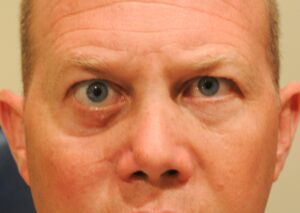
During the surgery by Dr. Fante, a custom, computer-generated 3D implant was used to reconstruct his cheekbone and eye socket using small, hidden incisions while he slept with general anesthesia. A graft was also used to support his lower eyelid properly.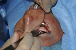
Several months after the surgery by Dr. Fante, you can see that his eyeball, eyelid, and cheek are each in a more normal position, with much better symmetry. The hidden incisions created almost no scar, and his eye was comfortable.
Who is a Good Candidate for Post-traumatic Facial Reconstruction?
There are several situations for which facial reconstructive surgery may be warranted. Common reasons for this process include:
- Trauma from an accident
- Cancer or other destructive diseases
- Severe infections, such as mucormycosis or necrotizing fasciitis (“flesh-eating bacteria”)
Any physical trauma to the face that results in eye injury, lacerations, soft tissue injury (including burns), and fractures to the jaw, nose, cheek, or eye socket may need to be treated with reconstructive surgery
How Are 3d Implants Used for Facial Reconstruction?
Severe trauma to the face can lead to permanent disfigurement. Facial implants have been used for years to restore patients’ natural features. These artificial structures are made with the most biologically inert materials, meaning that the body is much less likely to reject them. The objective of a 3D facial implant is to recreate the look, feel, and mechanics of real bone. 3D facial implants are revolutionary because they match specific anatomy, thereby improving patient outcomes. The use of 3D technology enables surgeons to observe the current and anticipated structure before stepping into the operating room.
How Are 3D Implants Created?
3D printing has been an extraordinary advancement in modern medicine. The utilization of computer-generated printing based on high-resolution digital imaging such as CT and MRI files, expands surgeons’ ability to address bone deficiencies and other consequences of traumatic facial injury using a customized approach.
Digital imaging builds the foundation on which a model of the patient’s undamaged bone structure can be made. This model is then transformed into a mirror image. From this mirror image model, a perfectly symmetrical facial prosthetic can be made. The 3D printed pieces are excellent for complex restoration, including use in the tiniest areas of the face, such as bone fragments.
Facial implants may be printed from titanium powder or porous polyethylene on laser machines. These machines heat the powder, fusing it into bone-like structures. The resulting implant represents a personalized prosthesis that can enable the patient to recognize themselves when they look in the mirror.
What is Involved in Facial Reconstruction Surgery?
The complexity of facial reconstruction surgery varies considerably based on the extensiveness and type of initial injury. Detailed imaging provides the necessary “road map” for optimal reconstruction, which may include:
- Rhinoplasty
- Orbital fracture repair using titanium screws, sheets, and plates
- Skin tissue transplant
- Bone grafting
- Facial implants
Surgery may be performed under general anesthesia in an accredited surgery center or hospital. Detailed surgical guides may be used to position incisions through which facial implants can be installed. Implants are situated directly over the bony structure to provide a sufficient base for soft tissue support. After positioning and observing the implant for correct contouring, incisions are closed with non-dissolvable or dissolvable sutures or a combination of the two.
Recovery After Post-traumatic Facial Reconstruction
Patients can expect to encounter noticeable bruising and swelling after facial reconstruction surgery. These side effects may last from 10 to 30 days. If non-dissolvable stitches have been used, they will be removed approximately one week after surgery. Comfort is supported with prescription pain medication and may be enhanced with cool compresses if approved by the surgeon.
After surgery, it is not uncommon for the face to look different than it did before the traumatic injury. Sometimes, multiple surgeries are needed to fully address the initial problem. Further improvement may be achieved with non-surgical treatments that minimize the appearance of scars. As noted above, additional treatment may be helpful years later.
We do our best to prepare each patient for the side effects and outcome of facial reconstruction based on their type of injury and techniques used during surgery.
Call Today for Post-traumatic Facial Reconstruction in Denver!
Anything can happen at any time. Luckily, there are options to correct traumas. Call (303) 839-1616 or fill out the contact us form on this page to schedule a consultation with facial plastic surgeon Dr. Robert Fante to learn more about post-traumatic facial reconstruction.
Oculofacial plastic surgery preop and postop instructions 2019
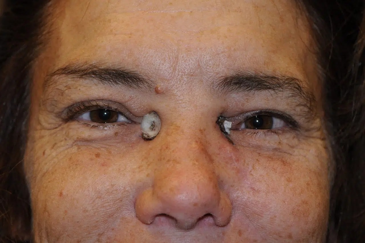
This woman was kicked by a horse and suffered extensive broken bones that changed the shape of her eye and made her cheekbone appear flat and low.
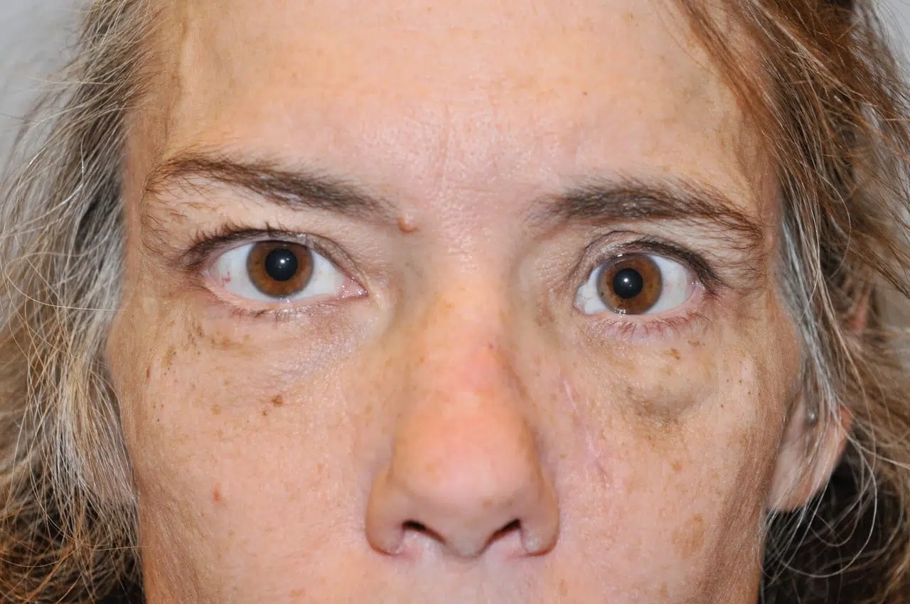
Before surgery by Dr. Fante, you can see that the inner corner of her eye is especially affected, making her eye appear shorter and her nose appear wider on the one side. This problem is called telecanthus. Her eye is also sunken, a condition known as enophthalmos.
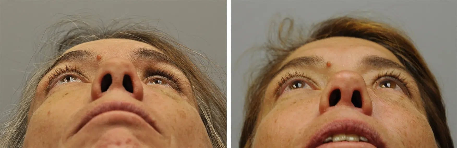
At surgery by Dr. Fante, a 3D custom, computer-generated implant was use to rebuild her cheekbone and eye socket, and a special wiring technique was used to anchor the inner corner of her eye. This photo shows her appearance at two weeks after surgery, and you can see bolsters still in place protecting the skin of the inner eye area.
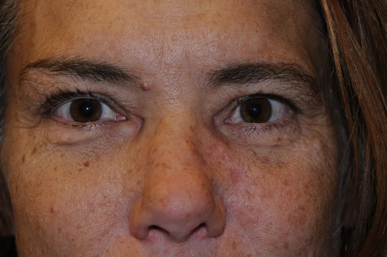
Finally, this photograph shows her appearance from the front after several months of healing. The set of photos taken from below shows that her sunken eye has been corrected.
This man had cancer of the sinus and cheekbone requiring surgery and radiation that left him with a sunken cheek and without a floor for his eye socket. As a consequence, his eye sank both down and back, causing double vision and obvious deformity.
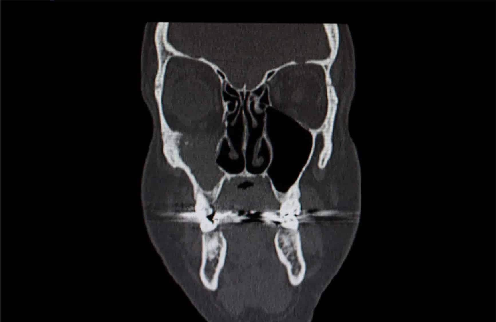
This coronal CT scan shows that the floor of his right eye socket (on your left as you look at it) is missing. His right eye has no support and is lower and deeper in his face than it should be.
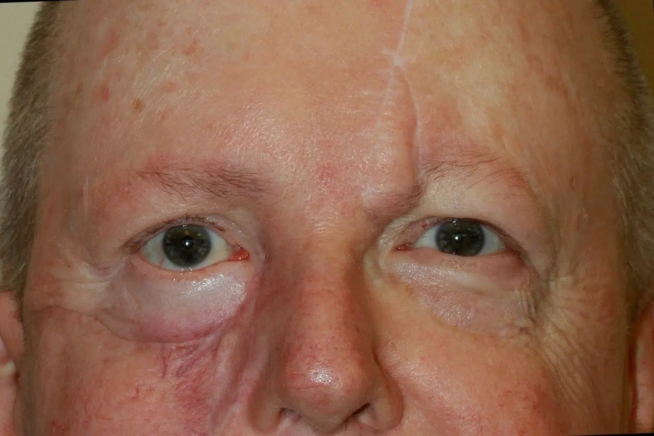
Surgery to repair his problems was complex and required both Dr. Fante and an ear, nose, and throat specialist. A new customized orbital (eye socket) floor was implanted, and his sinus and cheek were advanced and rebuilt. Here is his photo a few months later. Although not perfect, his eye was comfortable and his double vision had resolved.